Component PC · Provision of Clinical Dental Hygiene Services · 115 of 200 discipline-based items
NBDHE clinical services practice questions
29 original questions from the DHPrep NBDHE preview, with the correct answer and a rationale for every option.
Provision of Clinical Dental Hygiene Services is 115 of the 200 discipline-based items on the specifications in force, and 29 of the 50 free questions are tagged here. All 29 are printed below. Five of them ask you to interpret a synthetic teaching radiograph, and the image is on the page with the question.
What is here mixes standalone items with questions that share a patient profile. The profile includes the history and charting the question was written against. A case id that contains “MOCK” is still a practice item: it is not on either reserved mock form.
How to work this set. Mark the stage first: assess, plan, treat, or maintain. A periodontal procedure is the wrong key when the stem is still asking what the chart means. On a shared profile, use the numbers in the charting box, not a typical pocket depth you remember from class.
Planning and managing dental hygiene care
4 standalone questions in the free set.
Question 1
During a follow-up visit, an adult patient reports using mouthrinse daily, but still exhibits moderate gingival bleeding sites. What individualized educational strategy should the hygienist implement first?
- A. Encourage avoiding all spicy or acidic foods
- B. Evaluate and reinforce proper brushing and flossing techniquesCorrect answer
- C. Recommend switching to a stronger mouthrinse brand
- D. Suggest more frequent dental recall appointments
Answer
B. Evaluate and reinforce proper brushing and flossing techniques
Evaluating and reinforcing brushing and flossing ensures proper mechanical plaque removal, the primary method to address gingival bleeding.
Rationale
Mechanical plaque control is the primary preventive approach for gingival disease. Mouthrinses help, but if bleeding persists, technique or compliance with brushing and flossing is often deficient. Reinforcing these techniques addresses the root cause. The other options might be components of a long-term plan but do not address the likely deficiency in home care at this stage.
Why each option lands where it does
- A. Dietary advice may help but is not usually the primary cause of gingival bleeding.
- B (correct). Addresses the most common cause—insufficient or improper mechanical plaque removal.
- C. Stronger mouthrinse alone does not substitute for proper technique.
- D. Increasing recall frequency is not justified without first improving home care.
Decision rule. Select the strategy most directly addressing the likely cause of persistent symptoms.
Takeaway. Effective plaque removal through proper technique is essential for gingival health.
Common miss. Some believe mouthrinse alone is sufficient to control gingival disease.
Question 2
A patient with recurrent root caries and visible plaque claims their home care is sufficient. Which action best helps the hygienist assess and address the patient’s oral hygiene technique?
- A. Refer to periodontist
- B. Increase cleaning frequency
- C. Suggest antibacterial rinse
- D. Observe brushing techniqueCorrect answer
Answer
D. Observe brushing technique
Having the patient demonstrate their brushing technique uncovers misunderstandings and enables immediate, personalized feedback.
Rationale
Effective individualized education involves assessing actual patient technique rather than solely relying on self-report. By observing the patient demonstrate brushing, the hygienist can diagnose errors or misconceptions and provide tailored instruction. Increasing cleaning frequency or referrals doesn't address the underlying education need, and suggesting a rinse without engagement misses an opportunity to correct technique.
Why each option lands where it does
- A. Referral may be premature without first evaluating the patient’s current technique.
- B. More frequent cleanings do not identify or correct home care issues.
- C. Recommending a rinse does not address possible technique errors.
- D (correct). Direct observation reveals technique gaps and allows for targeted instruction.
Decision rule. Select options that involve assessment and patient engagement before escalating care.
Takeaway. Directly assessing patient technique is key for identifying and correcting oral hygiene gaps.
Common miss. Assuming patient self-reports are accurate without verification can miss skill deficits.
Question 3
During a dental hygiene appointment, a patient with type 1 diabetes mellitus reports sweating and lightheadedness. What is the FIRST action the dental hygienist should take?
- A. Activate emergency medical services
- B. Have the patient rinse with water
- C. Provide 100% oxygen via mask
- D. Administer oral glucose sourceCorrect answer
Answer
D. Administer oral glucose source
Symptoms of hypoglycemia should be treated immediately by administering a fast-acting oral glucose source as the first intervention.
Rationale
Sweating and lightheadedness in a diabetic patient suggest hypoglycemia, which is managed initially by giving glucose orally if the patient is conscious. Calling EMS is appropriate only if the patient becomes unresponsive. Rinsing with water does not address hypoglycemia. Oxygen is not indicated for hypoglycemia without respiratory compromise.
Why each option lands where it does
- A. EMS should be called if patient loses consciousness or does not improve, not as the first step.
- B. Rinsing does not resolve hypoglycemia and may delay proper treatment.
- C. Oxygen is not routinely indicated unless there is airway or breathing compromise.
- D (correct). Rapid glucose replaces deficit and addresses hypoglycemia symptoms immediately.
Decision rule. Identify patient consciousness and provide oral glucose as first-line therapy in hypoglycemia.
Takeaway. Prompt recognition and immediate action are critical in managing hypoglycemic episodes in diabetic patients.
Common miss. Students might over-prioritize EMS or oxygen before addressing the underlying cause in a conscious patient.
Question 4
A 60-year-old with controlled type 2 diabetes presents with 5–6 mm pockets, generalized bleeding on probing, and moderate attachment loss. Which dental hygiene intervention should be prioritized first to improve long-term periodontal health?
- A. Chlorhexidine rinse only
- B. Immediate referral for surgery
- C. Annual periodontal evaluation only
- D. Nonsurgical therapy and home care instructionCorrect answer
Answer
D. Nonsurgical therapy and home care instruction
Nonsurgical periodontal therapy and improved home care are the first-line approach for periodontitis, even in patients with diabetes.
Rationale
Current evidence supports initial nonsurgical therapy (scaling, root planing) along with personalized oral hygiene instruction for periodontitis, even in diabetic patients. Surgery or rinses alone do not address all etiologies and may not be immediately indicated without prior noninvasive measures.
Why each option lands where it does
- A. Chlorhexidine rinse alone is not sufficient to manage moderate periodontitis.
- B. Surgical referral is not indicated before nonsurgical therapy is attempted.
- C. Annual evaluation without intervention does not address active disease.
- D (correct). Nonsurgical therapy and home care are evidence-based first steps for periodontitis.
Decision rule. Select strategies that start with least invasive, evidence-based interventions appropriate for disease severity.
Takeaway. Initial nonsurgical therapy is the evidence-based strategy for managing periodontitis, even in medically compromised patients.
Common miss. Some may incorrectly assume surgery or rinses alone are preferred first steps in advanced cases.
Professional responsibility
1 standalone question in the free set.
Question 5
Which of the following best describes the principle of veracity in the dental hygiene code of ethics?
- A. Upholding justice in patient care
- B. Maintaining confidentiality of patient records
- C. Respecting the autonomy of the patient
- D. Communicating truthfully with patientsCorrect answer
Answer
D. Communicating truthfully with patients
Communicating truthfully with patients reflects the principle of veracity, which emphasizes honesty in professional relationships.
Rationale
Veracity refers to truthfulness and integrity in interactions between dental hygienists and patients. This principle requires practitioners to provide accurate information, avoid deception, and ensure patients are properly informed about their care. It differs from other ethical principles such as autonomy (respecting patient choices), confidentiality (protecting patient privacy), and justice (fair treatment of all patients). Recognizing these distinctions is essential for understanding and upholding professional responsibilities.
Why each option lands where it does
- A. Justice pertains to fairness and equality, not truthfulness.
- B. Confidentiality is a separate ethical principle focused on privacy, not veracity.
- C. Autonomy involves respecting patient decisions rather than specifically telling the truth.
- D (correct). Veracity specifically requires honesty and transparency with patients, aligning directly with truthful communication.
Decision rule. Select the option that most directly relates to truthfulness in the professional role.
Takeaway. Ethical principles guide professional conduct; veracity means being honest with patients.
Common miss. Candidates may confuse veracity with confidentiality or autonomy due to overlap in ethical terminology.
Case-based questions
A patient profile is reprinted with the charting the questions use. Only the free questions tagged to this component are on this page. A profile whose case id contains MOCK is still a practice item; it is not one of the reserved mock forms.
Patient profile 1
A 74-year-old male presents for a routine dental hygiene visit, reporting sensitivity to cold foods and occasional discomfort when brushing his teeth near the gumline. He lives independently but admits to having cut back on some self-care activities due to arthritis. He has a history of hypertension, controlled with medication, and recently began taking a calcium channel blocker. He has not smoked in over 30 years. Intraoral exam reveals several areas of gingival recession, with visible root surfaces and darkened cervical lesions on multiple posterior teeth. The patient’s oral hygiene appears inconsistent, and he reports brushing once daily with a medium-bristled manual toothbrush. He does not routinely use interdental cleaning devices or mouthrinse. His chief concern is preserving his remaining teeth and minimizing discomfort. He expresses mild dental anxiety, but is cooperative during the visit.
Vital Signs and Medical History
Blood pressure: 132/78 mmHg, Pulse: 72 bpm. PMH: Hypertension, osteoarthritis. Medications: Amlodipine 5 mg daily, Acetaminophen as needed. No drug allergies. Former smoker, quit at age 44. No diabetes or other systemic illness.
Periodontal Chart (Selected Teeth)
Teeth 3, 5, 14, 19, 30 (selected posterior teeth): - Probing depths (mm): 3-4 buccal, 2-3 lingual - Recession (mm): 2-4 buccal, 1-2 lingual - Bleeding on probing: Present at buccal of 3, 19, 30 - No mobility - Furcation: Class I involvement on 3, 14 - Plaque index: Moderate on exposed roots
Clinical Findings
Generalized gingival recession with visible root surfaces, especially teeth 3, 5, 19, and 30. Brownish, softened cervical lesions noted on buccal root surfaces of 3, 19, 30. No facial swelling. Slight cervical abrasions on several teeth. Mild dry mouth. No oral lesions or ulcers.
Full-Mouth Radiographic Survey
Periapical and bitewing radiographs show horizontal bone loss of 2-3 mm in posterior regions. Radiolucent lesions in the cervical root areas of 3, 19, and 30, with distinct loss of root surface density. No periapical radiolucencies, caries on coronal enamel, or large restorations.
Question 6
The patient’s exposed root surfaces and caries risk increase infection control demands. Which strategy is most appropriate to prevent cross-contamination during this hygiene appointment?
- A. Limit saliva ejector use
- B. Use a non-sterile prophy angle
- C. Rinse with baking soda solution
- D. Use disposable barrier coversCorrect answer
Answer
D. Use disposable barrier covers
Barriers prevent instrument and surface contamination during treatment.
Rationale
Disposable barriers protect equipment from contamination by saliva and debris during procedures on patients with exposed roots and potential bleeding. The other options do not adequately address cross-contamination or deviate from infection control guidelines.
Why each option lands where it does
- A. Properly used saliva ejectors protect the field; reduction offers no IPC benefit and may inconvenience patient.
- B. Prophy angles must be either sterilized or disposable, not non-sterile.
- C. Baking soda rinses lack evidence for infection control and do not prevent cross-transmission.
- D (correct). Barriers are required for infection control, especially with potential for contamination.
Decision rule. Apply evidence-based protocols for infection prevention.
Takeaway. Infection control relies on barriers, sterilization, and standard precautions.
Common miss. Believing cross-contamination is unlikely in hygiene undermines patient safety.
Patient profile 2
A 58-year-old man presents for a dental hygiene visit complaining of bleeding gums and occasional soreness when chewing. He has not seen a dentist in over three years due to concerns about cost and previous discomfort during cleanings. He reports brushing once daily but admits to rarely flossing. He was diagnosed with type 2 diabetes seven years ago and describes his blood sugar as “sometimes high.” He is a non-smoker and works as an accountant. He reports feeling anxious before dental appointments and requests to minimize discomfort during procedures. He also mentions feeling tired lately. Review of systems is otherwise unremarkable. Clinical evaluation is performed, and relevant findings are summarized in the following exhibits.
Medical and Dental History
Type 2 diabetes (diagnosed 7 years ago); latest HbA1c: 8.2% (last month). Metformin 1000 mg BID, Lisinopril 20 mg daily. Allergies: none reported. No tobacco, no alcohol use. Brushing 1x/day, flossing rarely. Dental visit >3 years ago. Reports anxiety about dental procedures and sensitivity to pain.
Vital Signs and Laboratory Values
Blood pressure: 138/88 mmHg; Pulse: 76 bpm; Temperature: 98.6°F. Capillary glucose (random): 182 mg/dL. HbA1c (last month): 8.2%.
Periodontal Charting (Selected Teeth)
Tooth 3: MB 6mm, B 5mm, DB 5mm, ML 5mm, L 5mm, DL 4mm; Tooth 8: MB 5mm, B 4mm, DB 4mm, ML 5mm, L 4mm, DL 4mm; Tooth 19: MB 7mm, B 6mm, DB 6mm, ML 6mm, L 6mm, DL 6mm. Generalized bleeding on probing. Several teeth with recession of 2-3mm.
Intraoral and Radiographic Findings
The gingiva is erythematous with loss of stippling and generalized swelling. Multiple posterior teeth exhibit moderate horizontal bone loss (30-40% root length). No evidence of periapical radiolucencies.
Question 7
Considering the patient’s infrequent flossing, elevated HbA1c, and concern about discomfort, which is the most effective individualized oral hygiene instruction to provide at this appointment?
- A. Demonstrate interdental brush useCorrect answer
- B. Instruct on toothpick use only
- C. Suggest brushing 3 times daily
- D. Recommend daily chlorhexidine rinse
Answer
A. Demonstrate interdental brush use
Demonstrating interdental brush use best addresses plaque removal for this patient’s periodontal status and dexterity.
Rationale
Interdental brushes are effective for cleaning larger interdental spaces created by attachment loss and recession, which are evident in this patient. Flossing may not be practical or effective in areas with periodontitis. Demonstrating proper brush use also addresses his anxiety by showing the process and allowing practice with feedback. While a chlorhexidine rinse may be used as adjunctive therapy, it does not replace mechanical plaque removal and should not be prescribed without clinical indication. Toothpick use is less effective and can traumatize tissues. Increasing brushing frequency is not as impactful for interdental areas.
Why each option lands where it does
- A (correct). Best option; matches clinical needs and supports manual dexterity for larger spaces.
- B. Toothpicks are less effective and may cause gingival trauma, especially in inflamed tissues.
- C. Increased brushing does little for interdental plaque; overbrushing may increase recession risk.
- D. Chlorhexidine should not replace mechanical cleaning; long-term use is not usually indicated without prescription.
Decision rule. Choose education matching clinical findings and the patient’s abilities.
Takeaway. Tailor hygiene education to individual needs and anatomy.
Common miss. Chlorhexidine rinses alone control periodontal disease. They are adjuncts, not substitutes.
Patient profile 3
A 27-year-old patient presents for her second dental hygiene visit during pregnancy. She is 24 weeks pregnant and reports recent bleeding from her gums during brushing, accompanied by mild tenderness. She brushes twice daily using a soft-bristled toothbrush and fluoridated toothpaste, and flosses about three times per week. She occasionally uses an alcohol-free mouthrinse. Her last dental visit was 6 months ago, prior to pregnancy, and she reports no previous periodontal treatment. She denies smoking, recreational drug use, or frequent snacking between meals but admits to occasional nausea and vomiting during the first trimester. Her medical history is significant for mild iron-deficiency anemia for which she takes prenatal vitamins and oral ferrous sulfate. She has no known drug allergies. She expresses concern about the safety of dental care during pregnancy and requests advice on maintaining oral health.
Medical and Dental History
Age: 27 years Sex: Female Pregnancy status: 24 weeks gestation (second trimester) Medications: Prenatal vitamin (contains folic acid, iron, calcium, vitamin D), Ferrous sulfate 325 mg daily Allergies: None Medical conditions: Mild iron-deficiency anemia Tobacco use: None Alcohol use: None Dental history: Regular dental care, last visit 6 months ago, no prior periodontal treatment
Vital Signs and Clinical Findings
Blood pressure: 110/68 mm Hg Heart rate: 84 bpm Respiratory rate: 16 Temperature: 98.6°F Height: 5'5" Weight: 153 lbs Extraoral exam: No abnormalities Intraoral soft tissue: Generalized erythema and edema of marginal and papillary gingiva, especially maxillary anterior region. Bleeding on gentle probing, no ulcerations. Dental plaque moderate, calculus minimal.
Periodontal Probing Chart (Selected Teeth, 6 Sites/tooth)
Tooth 6: 3-4-3 (B), 2-3-2 (L) Tooth 8: 4-4-3 (B), 2-3-2 (L) Tooth 9: 4-3-4 (B), 2-2-3 (L) Tooth 11: 3-3-3 (B), 2-2-2 (L) Bleeding on probing at all sites listed. No clinical attachment loss. Pseudopocketing likely present.
Radiographic Findings
Bitewing radiographs show no detectable crestal bone loss. No interproximal caries. Interdental septa appear intact. Calculus is not visible radiographically.
Question 8
A 28-year-old pregnant woman presents with moderate gingival inflammation and plaque accumulation. Which oral hygiene topic should be emphasized during today’s appointment?
- A. Importance of daily flossingCorrect answer
- B. Timing of elective dental surgery
- C. Risks of over-the-counter pain medications
- D. Safe use of topical anesthetics
Answer
A. Importance of daily flossing
Education should focus on improving daily flossing to control plaque and reduce gingival inflammation.
Rationale
Her current hygiene routine is good but could be improved by increasing flossing frequency, which directly addresses plaque control and gingival bleeding. Surgery and anesthetic topics are less immediate, and pain medication risks, while important, are not as relevant since she has not reported pain or use.
Why each option lands where it does
- A (correct). Daily flossing is essential to reduce plaque and manage pregnancy-associated gingival inflammation.
- B. Elective dental surgery timing is not relevant to her current oral findings or preventive needs.
- C. Pain medication risks are not the main priority without reported pain or medication use.
- D. Topical anesthetics are not indicated for routine preventive care and are not the primary concern for this visit.
Decision rule. Prioritize modifiable risk factors in individualized patient education.
Takeaway. Daily flossing is critical to control pregnancy-associated gingival inflammation.
Common miss. Candidates may prioritize procedural or medication topics over daily home care.
Patient profile 4
A 46-year-old male presents for a dental hygiene appointment after a five-year absence from professional care. The patient admits to avoiding dental visits due to severe dental anxiety and a pronounced gag reflex, which he says make dental procedures 'nearly impossible' without significant distress. He brushes twice daily but finds flossing difficult due to his gagging tendency. He expresses concern about 'bleeding gums' and 'sensitive teeth.' The patient is generally healthy, tries to control his anxiety with breathing exercises, and recently began taking sertraline for depression. He does not smoke or use alcohol. On examination, the patient appears anxious and is visibly tense during all intraoral procedures, with a pronounced gag response elicited during attempted intraoral radiography. He reports no known drug allergies. In your role as the dental hygienist, you must assess and plan appropriate follow-up and interventions to address his oral health and psychosocial needs.
Exhibit 1: Medical and Dental History
Medical conditions: Generalized anxiety disorder, depression Current medications: Sertraline 50 mg daily (started 6 weeks ago) Allergies: None Smoking/alcohol: None Last dental visit: 5 years ago At-home care: Brushes 2x daily with fluoride toothpaste, rarely flosses due to gagging Reports: Bleeding gums, tooth sensitivity, severe dental anxiety, strong gag reflex Family history: No significant history relevant to oral health
Exhibit 2: Vital Signs and Clinical Findings
Blood pressure: 134/82 mmHg Pulse: 88 bpm Respiratory rate: 18 breaths/min Temperature: 98.2°F Appearance: Alert, anxious, fidgeting Extraoral: No lesions, mild tension in jaw Intraoral: Generalized moderate plaque accumulation, marginal gingival inflammation, mild recession on 7-10, palatal and posterior surfaces elicit strong gag reflex
Exhibit 3: Periodontal Chart (Teeth 3, 8-10, 14, 19, 24-26, 30)
Tooth | MB | B | DB | DL | L | ML | Furcation 3 | 5 | 4 | 5 | 4 | 4 | 4 | II 8 | 3 | 3 | 4 | 3 | 3 | 3 | - 9 | 3 | 4 | 4 | 4 | 4 | 3 | - 10 | 3 | 4 | 4 | 3 | 4 | 3 | - 14 | 4 | 4 | 5 | 5 | 4 | 4 | I 19 | 4 | 4 | 4 | 4 | 3 | 3 | II 24 | 3 | 4 | 4 | 3 | 3 | 3 | - 25 | 3 | 3 | 4 | 3 | 3 | 3 | - 26 | 3 | 4 | 4 | 3 | 4 | 3 | - 30 | 4 | 4 | 5 | 4 | 4 | 4 | II Bleeding on probing: generalized Mobility: 1 (teeth 3, 14, 19, 30)
Exhibit 4: Radiographic Findings
Bitewing and limited periapical radiographs (only partially completed due to gagging): show generalized horizontal bone loss (25-40%), no periapical pathology, moderate calculus deposits, no visible caries. No evidence of root fractures or periapical abscesses.
Question 9
A 55-year-old patient presents with generalized plaque accumulation and reports increased root sensitivity. Which preventive measure would best address both his caries risk and sensitivity at home?
- A. High-fluoride toothpaste useCorrect answer
- B. Plaque-disclosing tablets only
- C. Monthly in-office fluoride varnish
- D. Daily chlorhexidine rinse
Answer
A. High-fluoride toothpaste use
High-fluoride toothpaste addresses root sensitivity and caries risk with manageable home use.
Rationale
This patient has exposed root surfaces (mild recession), sensitivity, and moderate plaque. High-fluoride toothpaste is effective for reducing sensitivity and caries risk, and is well tolerated for home use. Chlorhexidine rinses are effective against plaque, but long-term use has adverse effects and may not address sensitivity. In-office varnish is useful, but as a monthly only intervention it is less impactful than daily home care. Disclosing tablets help with motivation but do not directly prevent caries or sensitivity.
Why each option lands where it does
- A (correct). High-fluoride toothpaste offers daily home application, reducing caries risk and sensitivity effectively.
- B. Plaque-disclosing tablets help with plaque identification but do not address sensitivity or remineralization.
- C. In-office fluoride varnish is beneficial but lacks the frequency of home fluoride exposure.
- D. Chlorhexidine rinse targets biofilm but does not reduce sensitivity or provide fluoride.
Decision rule. Choose agents that are both effective and compatible with patient's self-care ability.
Takeaway. Daily home fluoride use reduces sensitivity and risk.
Common miss. Chlorhexidine is not used long-term for sensitivity.
Patient profile 5
A 55-year-old male presents for a dental hygiene visit. He reports that his last dental cleaning was two years ago. He has noticed gum bleeding while brushing and occasional bad breath. He is concerned about loose teeth in the back of his mouth and mentions that his gums have been receding. He has a 15-year history of type 2 diabetes, managed with oral medication, and states his blood sugar "isn't always under control." He denies tobacco or recreational drug use. His current medications include metformin and lisinopril. The patient’s physician recently reported an HbA1c of 8.4%. Review of systems is negative for other significant findings. Clinical examination and radiographs are performed. Relevant data are provided in the exhibits.
Vital Signs and Medication List
Blood pressure: 138/87 mm Hg; Pulse: 82 bpm; Respiratory rate: 16 breaths/minute; Temperature: 98.7°F. Medications: - Metformin 1000 mg PO BID - Lisinopril 10 mg PO daily
Medical Laboratory Results
Hemoglobin A1c: 8.4% (target <7.0%) Fasting glucose: 166 mg/dL (normal 70–99 mg/dL) Serum creatinine: 1.0 mg/dL (normal) Urinalysis: Negative for protein
Periodontal Chart (selected teeth: 2, 3, 14, 19, 30, 31)
Tooth 2: Probing depths 5-7 mm mesiobuccal/distobuccal; bleeding on probing (BOP) all sites Tooth 3: Probing depths 6-7 mm buccal, 4-5 mm lingual; BOP Tooth 14: Probing depths 5-7 mm; BOP Tooth 19: Probing depths 4-6 mm; BOP Tooth 30: Probing depths 6-7 mm; BOP; Class II mobility Tooth 31: Probing depths 5-6 mm; BOP
Full-Mouth Intraoral Findings
Heavy supragingival and subgingival calculus, generalized moderate plaque, gingival erythema and edema, recession 2-4 mm on molars, spontaneous bleeding in posterior regions, halitosis.
Posterior Bitewing Radiograph (Described)
Bitewing radiograph shows generalized horizontal bone loss on all posterior teeth; 30–40% root length remaining for teeth 2, 3, 14, 19, 30, and 31. No carious lesions visible.
Question 10
A 52-year-old patient with poorly controlled type 2 diabetes presents for periodontal therapy. Which supportive service will most effectively help the patient improve glycemic control and lower periodontal risk?
- A. Referral to a certified diabetes educatorCorrect answer
- B. Referral for orthodontic evaluation
- C. Scheduling for dental sealant placement
- D. Recommendation for fluoride varnish application
Answer
A. Referral to a certified diabetes educator
Referral to a diabetes educator supports patients in improving glycemic control, reducing periodontal and systemic complications.
Rationale
Poorly controlled diabetes is a key modifiable risk factor for periodontitis. Certified diabetes educators can work with patients on dietary strategies, medication adherence, and blood glucose monitoring. Improved glycemic control will improve healing, reduce periodontal inflammation, and decrease long-term complications. Orthodontic evaluation, fluoride varnish, and sealants are not directly relevant to his systemic risk or periodontal stability.
Why each option lands where it does
- A (correct). A certified diabetes educator provides guidance on blood glucose management, which is critical for periodontal health in diabetic patients.
- B. Orthodontic evaluation does not address glycemic control or reduce periodontal risk in diabetic patients.
- C. Dental sealants are used to prevent caries, not to manage glycemic control or periodontal risk.
- D. Fluoride varnish helps prevent caries but does not impact glycemic control or periodontal disease risk.
Decision rule. Refer medically complex patients for relevant systemic disease education/support.
Takeaway. Improving glycemic control is vital for successful periodontal outcomes in diabetic patients.
Common miss. Neglecting the link between systemic health support and oral disease management is a common error.
Question 11
What is the most significant periodontal finding for teeth 2, 3, 14, 19, 30, and 31, supporting active disease in this patient?
- A. Absence of caries on these teeth
- B. Generalized mild recession only
- C. Bleeding on probing and deep pocketsCorrect answer
- D. Occasional plaque and calculus
Answer
C. Bleeding on probing and deep pockets
Bleeding on probing with 5–7 mm pockets is the strongest indicator of active periodontitis.
Rationale
The combination of deep probing depths and BOP on multiple posterior teeth reflects ongoing periodontal breakdown. Plaque and calculus, while important risk factors, are not definitive evidence of current disease activity. Caries status is unrelated to periodontal status. Mild recession alone can occur in healthy but older patients.
Why each option lands where it does
- A. Caries status does not inform about periodontal disease.
- B. Mild recession may be physiologic or post-inflammatory, not disease activity per se.
- C (correct). Deep pockets plus BOP confirm current inflammation and connective tissue breakdown.
- D. Plaque/calculus presence is not diagnostic of disease activity.
Decision rule. Prioritize BOP and pockets when evaluating periodontal status.
Takeaway. Deep pockets with BOP are cornerstones of diagnosing active periodontitis.
Common miss. Relying on non-specific findings (like recession) can mask active disease.
Patient profile 6
A 41-year-old male presents for a dental hygiene appointment after a five-year hiatus from care. He expresses significant dental anxiety, reporting previous negative experiences and an intense fear of dental instruments. He admits to a strong gag reflex, especially during dental x-rays and intraoral procedures. The patient is otherwise communicative and appears alert but anxious. He reports brushing once daily and rarely flossing due to discomfort and fear of triggering his gag reflex. He brings a medication list and shares recent vital signs from his primary care physician. He denies known allergies but reports avoiding routine dental care due to fear. His chief concern is 'bleeding gums and bad breath.'
Medical and Dental History
Relevant medical history: History of hypertension, controlled. No diabetes. No cardiac events or surgeries. Dental history: Avoidance of care for 5 years, prior dental anxiety, reports significant gagging during dental x-rays. Medications: Lisinopril 20 mg daily. No allergies. Tobacco: Never. Last dental visit: 5 years ago. Family history: Non-contributory.
Vital Signs
Blood pressure: 138/86 mmHg Pulse: 85 bpm Respiratory rate: 15/min Temperature: 98.1°F BMI: 29
Partial Periodontal Chart (teeth 2, 3, 6, 8, 9, 14, 18, 19, 25, 30, 31)
Tooth 2: 4/5/4 mm, 2/3/3 mm, BOP Tooth 3: 4/5/5 mm, 3/4/5 mm, BOP Tooth 6: 3/3/3 mm, 2/2/2 mm, no BOP Tooth 8: 2/2/3 mm, 2/2/2 mm, BOP Tooth 9: 2/3/3 mm, 2/2/3 mm, BOP Tooth 14: 5/6/5 mm, 4/5/6 mm, BOP, recession 2 mm Tooth 18: 3/4/4 mm, 2/3/3 mm, BOP Tooth 19: 4/5/5 mm, 3/4/4 mm, BOP Tooth 25: 2/2/3 mm, 2/2/2 mm, no BOP Tooth 30: 4/5/4 mm, 3/5/4 mm, BOP Tooth 31: 4/4/4 mm, 3/3/4 mm, BOP
Soft Tissue and Head & Neck Exam
Extraoral: No lymphadenopathy, TMJ non-tender, no lesions. Intraoral: Palate and oropharynx WNL. Slight erythema of gingiva, especially posterior. Tongue: WNL. Floor of mouth: WNL. Multiple areas of mild plaque and calculus. No suspicious oral lesions.
Descriptive Note: Periapical Radiograph Attempt
During attempted posterior periapical x-rays, the patient became visibly distressed and began gagging. The radiograph could not be completed; only anterior bitewings were obtained successfully, showing moderate interproximal bone loss on posterior teeth.
Question 12
Refer to the bitewing radiograph provided. Which tooth displays the greatest extent of interproximal alveolar bone loss as demonstrated in the image?
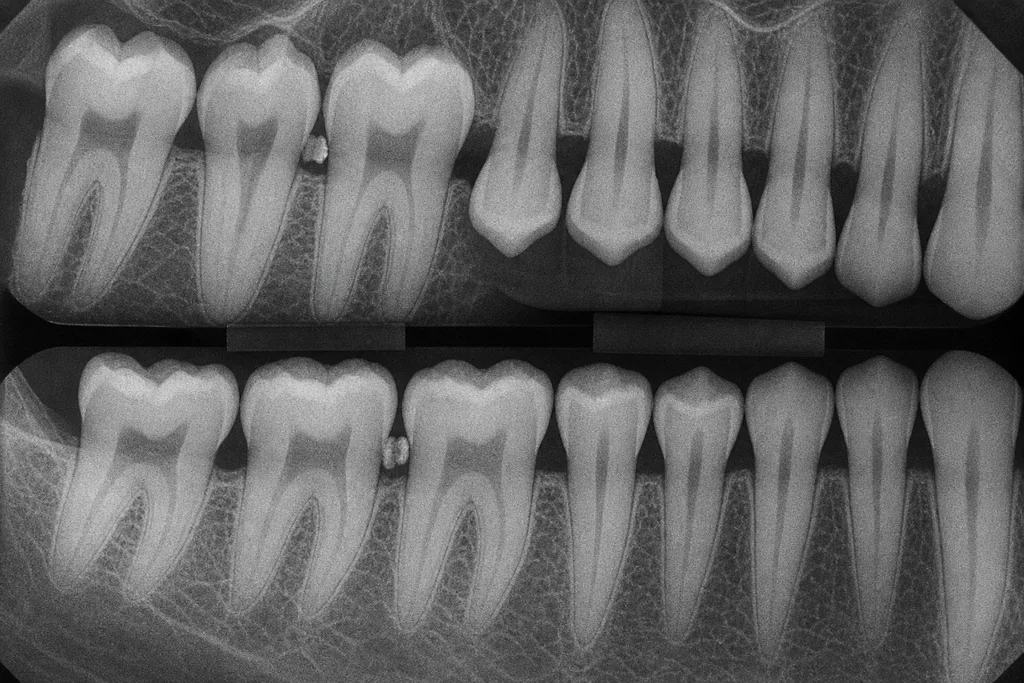
- A. Tooth 19 mesial
- B. Tooth 3 distal
- C. Tooth 14 mesialCorrect answer
- D. Tooth 31 distal
Answer
C. Tooth 14 mesial
Tooth 14 mesial displays the greatest interproximal bone loss on the radiograph, extending apically beyond the CEJ relative to other sites.
Rationale
The candidate must evaluate the image for crestal bone height relative to the cementoenamel junction (CEJ). Tooth 14 mesial clearly shows greater vertical bone loss compared with other interproximal surfaces. This is corroborated by severe clinical probing depths and recession at that site, but the answer relies entirely on the radiographic appearance. The other options represent sites with mild to moderate loss, but not to the same extent visible at tooth 14 mesial.
Why each option lands where it does
- A. Incorrect. While moderate bone loss is present, it does not reach the severity seen on tooth 14 mesial.
- B. Incorrect. Some bone loss is present, but it is less extensive than on tooth 14 mesial.
- C (correct). Correct. The radiograph reveals pronounced bone loss at tooth 14 mesial, which is more severe than at other listed sites.
- D. Incorrect. This site exhibits mild to moderate loss, not the most extensive in the image.
Decision rule. Select the site with the greatest vertical distance from the CEJ to the remaining alveolar crest on interproximal surfaces.
Takeaway. Interpretation of radiographs is essential for identifying the most severely affected periodontal sites.
Common miss. Students may incorrectly assume clinical measurements determine the answer, but only the radiograph is relevant here.
Patient profile 7
A 15-year-old male comes to the dental hygiene clinic for a routine checkup. His mother is present and reports that he has been increasingly self-conscious about his smile. He brushes once daily and flosses occasionally. He admits to drinking several cans of soda per day and enjoys candy frequently. He has not visited the dentist in the past two years. He denies any medical conditions. During the exam, he is cooperative but anxious. There is no history of allergies. The patient takes no medications and reports no recent illnesses or hospitalizations. He reports mild, intermittent sensitivity to cold in the upper right molar area but denies spontaneous pain. He has not noticed any swelling, bleeding, or loose teeth. You proceed with a comprehensive oral evaluation, including periodontal and radiographic assessments.
Vital Signs and Medical History
Blood pressure: 110/72 mmHg Heart rate: 80 bpm Respiratory rate: 16/min Temperature: 98.2°F Medications: None Allergies: None Medical conditions: None reported
Dental History and Reported Habits
Dental visits: Last visit 2 years ago Oral hygiene: Brushes once daily, flosses occasionally Diet: 3–4 cans of soda daily, frequent candy intake, snacks between meals Chief concern: Self-conscious about smile, mild cold sensitivity in upper right molar
Head, Neck, and Intraoral Examination
Extraoral: No lymphadenopathy or swelling TMJ: No clicking or deviation Intraoral: No lesions or abnormal findings on mucosa, tongue, or floor of mouth Occlusion: Mild crowding lower anteriors
Periodontal Chart (Selected Teeth)
Tooth 2: 2, 3, 2 mm B | 2, 2, 3 mm L, no BOP Tooth 3: 3, 4, 3 mm B | 2, 4, 3 mm L, BOP on mesial Tooth 8: 2, 2, 2 mm B | 2, 2, 2 mm L, no BOP Tooth 14: 2, 2, 2 mm B | 3, 3, 2 mm L, no BOP Tooth 19: 3, 3, 3 mm B | 3, 4, 3 mm L, no BOP Tooth 30: 2, 2, 3 mm B | 2, 2, 3 mm L, BOP on distal General: No recession, no furcation, minimal calculus, moderate plaque
Bitewing Radiograph Findings
Bitewing radiographs show radiolucencies at the cervical third of the distal surface of tooth 3 and the occlusal surface of tooth 14. No evidence of periapical pathology. Interdental crests appear intact.
Question 13
The intraoral exam shows mild crowding of the lower anteriors, moderate plaque, and no abnormal mucosal lesions. Based on these findings, what is the priority oral evaluation concern?
- A. Screening for oral cancer
- B. Urgency of soft tissue biopsy
- C. Need for orthodontic referral
- D. Caries risk from moderate plaqueCorrect answer
Answer
D. Caries risk from moderate plaque
The main concern is caries risk from visible moderate plaque.
Rationale
With active caries, high sugar intake, and moderate plaque, caries management is the primary concern here. Mild crowding may eventually warrant orthodontic monitoring, but is not urgent. There are no suspicious mucosal findings, so oral cancer screening and biopsy are not indicated.
Why each option lands where it does
- A. No lesions identified to raise suspicion of malignancy.
- B. No lesion to biopsy based on the exam.
- C. Mild crowding is not causing urgent functional or esthetic problems.
- D (correct). Plaque directly contributes to risk for further caries, making intervention critical.
Decision rule. Prioritize the most urgent, modifiable risk to oral health.
Takeaway. Active plaque and caries risk take priority for intervention.
Common miss. Do not recommend biopsy without a clinical indication.
Question 14
Your head and neck examination reveals no lymphadenopathy, swelling, or TMJ abnormalities. What is the most clinically appropriate next step given these findings?
- A. Document negative findings only
- B. Order blood tests for infection
- C. Proceed with intraoral examCorrect answer
- D. Refer for MRI evaluation
Answer
C. Proceed with intraoral exam
Normal head and neck findings allow you to proceed with intraoral assessment.
Rationale
With unremarkable head and neck results (no swelling, node enlargement, or TMJ disorder), the proper clinical sequence is to move on to the intraoral exam. Advanced imaging or lab studies are unnecessary in the absence of signs or symptoms. Documentation is vital but never substitutes for the completion of the examination process.
Why each option lands where it does
- A. Documentation alone is insufficient; the exam should continue.
- B. Blood work is for suspected systemic or infectious disease, not supported here.
- C (correct). Progressing to intraoral examination follows a logical, systematic approach.
- D. MRI is reserved for suspected soft tissue or TMJ pathology, not indicated by findings.
Decision rule. Complete patient assessment systematically before referrals or special tests.
Takeaway. A negative head and neck exam is followed by intraoral evaluation.
Common miss. Imaging or labs are not routine in the absence of pathology.
Patient profile 8
A 54-year-old male presents for a dental hygiene recall appointment. He reports that he has been coming regularly for professional cleanings. The patient mentions persistent fatigue, a metallic taste in his mouth, and easy bruising. He undergoes hemodialysis three times weekly. He states he received dialysis the day before this appointment. His chief concern is maintaining his oral health while managing his overall medical condition. The review of systems is notable for hypertension and generalized bone/joint discomfort. The patient expresses mild dental anxiety, especially regarding discomfort during procedures. He does not report any known allergies. The patient brings a recent medication list and laboratory results from his medical provider. Clinical and radiographic examination reveals moderate plaque accumulation, generalized edema, and evidence of periodontal bone loss. You have access to his medication list, selected laboratory values, and a portion of his periodontal chart.
Medication List
1. Lisinopril 20 mg daily 2. Amlodipine 5 mg daily 3. Sevelamer 800 mg with meals 4. Epoetin alfa injection during dialysis sessions 5. Vitamin D supplement, 1000 IU daily No known drug allergies
Recent Laboratory Values
Blood Urea Nitrogen (BUN): 60 mg/dL (↑) Serum Creatinine: 7.1 mg/dL (↑) Platelet Count: 120,000/μL (mildly ↓) Calcium: 7.8 mg/dL (↓) Phosphorus: 5.3 mg/dL (↑) Hemoglobin: 10.2 g/dL (↓)
Selected Vital Signs
Blood Pressure: 142/86 mm Hg Heart Rate: 86 bpm Respiratory Rate: 16/min Temperature: 98.2°F
Partial Periodontal Charting (Teeth #3, #14, #19, #30)
Tooth #3: MB 5, B 4, DB 4, DL 5, L 4, ML 3; BOP all sites Tooth #14: MB 6, B 5, DB 6, DL 6, L 5, ML 4; BOP all sites Tooth #19: MB 5, B 4, DB 5, DL 4, L 5, ML 4; BOP all sites Tooth #30: MB 4, B 4, DB 5, DL 4, L 4, ML 3; BOP all sites
Extraoral Image Description
The patient’s arms show several healed puncture marks consistent with vascular access for hemodialysis. Mild pallor is visible. No facial swelling or asymmetry noted.
Question 15
A 52-year-old patient returns three months after scaling and root planing. Which finding most strongly supports scheduling additional periodontal maintenance visits?
- A. Persistence of bleeding on probingCorrect answer
- B. No change in medication list
- C. Stable vital signs at appointments
- D. Absence of dental calculus
Answer
A. Persistence of bleeding on probing
Ongoing bleeding on probing signifies a continued need for maintenance and possible retreatment.
Rationale
Bleeding on probing after therapy suggests persistent inflammation and ongoing periodontal disease activity. Frequent maintenance is needed to control disease and prevent recurrence. Stable vital signs, absence of calculus, or medication stability do not specifically guide periodontal maintenance scheduling.
Why each option lands where it does
- A (correct). Bleeding on probing reflects active inflammation, indicating the need for continued maintenance.
- B. Medication changes are important but do not directly indicate periodontal disease activity.
- C. Vital signs are general health indicators, not specific to periodontal status.
- D. Calculus absence is positive, but inflammation may still persist without it.
Decision rule. Look for clinical evidence of ongoing disease to guide follow-up.
Takeaway. Bleeding on probing guides maintenance needs after therapy.
Common miss. Maintenance frequency should be based on inflammation, not only calculus or systemic factors.
Patient profile 9
A 46-year-old male presents for a dental hygiene appointment. He reports sensitivity in his lower molars, occasional bleeding while brushing, and persistent halitosis. He has a 25-year history of daily cigarette smoking (1.5 packs/day) and consumes 2 beers most evenings. The patient last visited a dentist 18 months ago and reports having had several deep cleanings in the past. He has no known drug allergies and takes lisinopril for hypertension, which is controlled. Blood pressure at today’s appointment is 134/86 mmHg, pulse is 80 bpm. His medical history is otherwise noncontributory. The patient appears anxious but is cooperative. Clinically, generalized plaque accumulation, moderate to heavy subgingival calculus, and recession are noted, along with tooth mobility. The attached periodontal chart and radiographic summary are available for review.
Medication List
1. Lisinopril 20 mg, once daily (for hypertension) 2. No other prescription or OTC medications 3. No known drug allergies
Vital Signs
Blood pressure: 134/86 mmHg Pulse: 80 bpm Respirations: 16/min Temperature: 98.2°F
Periodontal Chart (Partial)
Teeth: 18-21, 29-31 Probing depths (mm): #18: MB 6, B 5, DB 5 | ML 6, L 5, DL 4 #19: MB 5, B 6, DB 6 | ML 5, L 6, DL 5 #20: MB 6, B 5, DB 6 | ML 5, L 5, DL 5 #21: MB 4, B 4, DB 4 | ML 5, L 4, DL 4 #29: MB 6, B 6, DB 5 | ML 6, L 6, DL 5 #30: MB 7, B 8, DB 6 | ML 6, L 7, DL 6 #31: MB 5, B 5, DB 5 | ML 6, L 5, DL 4 Tooth mobility: #19 (Class II), #30 (Class II), #20 (Class I) Gingival recession: 2-4 mm on all lower molars
Radiographic Summary
A full-mouth series shows generalized horizontal bone loss of 40-60% in posterior regions, vertical bone defects at #19 and #30, widened periodontal ligament spaces, and calculus deposits on root surfaces of lower molars. No carious lesions or periapical pathology are evident.
Question 16
Reviewing the bitewing radiograph, on which set of teeth is the horizontal bone loss most extensive, and how much of the root length does it approximately involve?
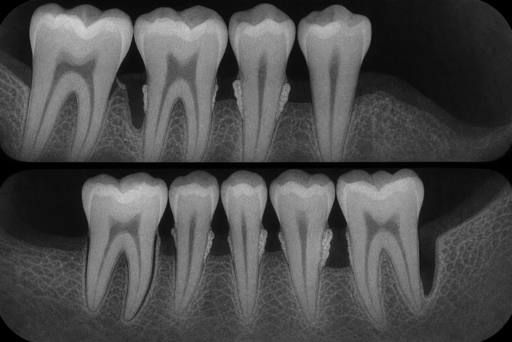
- A. Teeth 18-21, 40-60%Correct answer
- B. Teeth 29-31, 20-30%
- C. Teeth 18-21, 70-80%
- D. Teeth 22-27, 10-20%
Answer
A. Teeth 18-21, 40-60%
The most extensive horizontal bone loss involves teeth 18-21, affecting approximately 40-60% of the root length.
Rationale
On the radiograph, teeth 18-21 demonstrate generalized horizontal bone loss consistent with 40-60% of the root structure lost, with the alveolar crest notably closer to the mid-root than the CEJ. The same degree of bone loss is not seen in teeth 29-31, and anterior teeth (22-27) do not display significant bone loss. The option of 70-80% is an overestimation, as the radiograph does not show bone loss reaching near the apex.
Why each option lands where it does
- A (correct). Teeth 18-21 show clear horizontal bone loss of 40-60% of the root, as measured from CEJ to apex.
- B. Teeth 29-31 have similar bone loss, but the percentage given is an underestimation for this case.
- C. 70-80% bone loss would approach the apex, which is not evident on the radiograph for 18-21.
- D. Teeth 22-27 are not involved; anterior teeth retain normal bone levels in this image.
Decision rule. Use root length as reference to visually estimate the proportion of bone loss, focusing on posterior regions per description.
Takeaway. Assessing horizontal bone loss requires measuring from the CEJ and estimating the percentage of root affected on radiographs.
Common miss. Candidates may underestimate or overestimate bone loss by not properly using anatomical landmarks on radiographs.
Patient profile 10
A 43-year-old male presents for a dental hygiene appointment, reporting increased gum bleeding and discomfort over the past several months. He last saw a dentist one year ago. His medical history is significant for a chronic condition managed by an infectious disease specialist. He takes regular medications but denies experiencing dry mouth or recent hospitalizations. He reports a 20-year history of smoking, but quit five years ago. His oral hygiene routine includes brushing twice daily and occasional flossing. On intraoral examination, you note generalized erythema and swelling of the gingiva, along with bleeding on probing and significant plaque accumulation. The patient expresses concern about his oral health and asks about ways to improve his gum condition. Radiographs indicate notable bone loss, particularly in the molar and premolar regions.
Medical History and Medication List
• Diagnosed HIV infection, CD4+ count: 475 cells/mm3 (last checkup, 3 months ago) • Viral load: undetectable • Medications: Emtricitabine/tenofovir 200/300 mg daily, Dolutegravir 50 mg daily • No medication allergies • Smoked 20 pack-years, quit 5 years ago • No diabetes or cardiovascular disease • Last dental visit: 1 year ago • Vital signs today: BP 122/80 mmHg, Pulse 78 bpm, Temp 98.7°F
Dental Findings and Periodontal Chart
• Generalized gingival erythema and edema • Bleeding on probing: >50% of sites • Plaque index: 2.5/3 • Probing depths (selected): - #2: MB 5, B 6, DB 4, ML 5, L 6, DL 5 - #3: MB 7, B 6, DB 7, ML 6, L 7, DL 5 - #6: MB 4, B 4, DB 3, ML 3, L 3, DL 2 - #8: all sites 2-3 - #14: MB 7, B 8, DB 7, ML 7, L 8, DL 6 - #19: MB 6, B 6, DB 5, ML 5, L 5, DL 4 - Generalized CAL 3-6 mm • Mobility: #14 and #3, grade II • Furcation involvement: #3 buccal and #14 mesial, grade II
Laboratory Testing Summary
• WBC: 5,200/mm3 (reference: 4,000–11,000) • Platelets: 227,000/mm3 (reference: 150,000–400,000) • Hemoglobin: 13.7 g/dL • CD4+ T-cell count: 475 cells/mm3 • HIV viral load: undetectable • No other abnormalities noted
Radiographic Findings
Full-mouth periapical and bitewing radiographs demonstrate generalized horizontal bone loss in posterior regions, with localized angular defects at #3, #14, and #19. No periapical radiolucencies or carious lesions observed.
Question 17
At the four-week reassessment following initial therapy, what clinical parameter is the most valuable indicator for evaluating the success of periodontal therapy in this patient?
- A. Amount of dental calculus present
- B. Patient self-reported comfort
- C. Reduction in bleeding on probingCorrect answer
- D. Change in tooth color
Answer
C. Reduction in bleeding on probing
Reduction in bleeding on probing is the key, objective measure of improved periodontal health after therapy.
Rationale
Bleeding on probing (BOP) is a sensitive and objective marker of active inflammation and is routinely used to monitor periodontal treatment response. Decreased BOP rates after therapy indicate effective resolution of inflammation. Change in tooth color is not a clinical endpoint of therapy. Patient comfort is important but subjective and may not reflect disease status. Amount of calculus is less critical in short-term reevaluation than inflammatory signs.
Why each option lands where it does
- A. Residual calculus is less significant than reduction of inflammation in short-term reassessment.
- B. Self-reported comfort does not objectively measure periodontal health.
- C (correct). BOP is the gold standard for assessing active periodontal disease and treatment outcome.
- D. Tooth color does not reflect periodontal status or healing.
Decision rule. Use objective inflammatory markers—especially BOP—to evaluate periodontal therapy.
Takeaway. Monitor bleeding on probing at reassessment to gauge treatment effectiveness.
Common miss. Confusing patient comfort with objective clinical success may miss ongoing disease.
Patient profile 11
A 44-year-old man presents for a dental hygiene appointment, reporting issues with bleeding gums and recent challenges maintaining oral hygiene due to swelling of the gum tissue. He reports a long-standing seizure disorder, well-controlled with medication, and no known allergies. His last seizure was over a year ago. He has not smoked for ten years and drinks alcohol only occasionally. Medical history includes high blood pressure and a generalized anxiety disorder. The patient lives independently, works as an accountant, and expresses some concern about his appearance and oral health. He brushes twice a day, struggles to floss due to gum enlargement, and last visited the dentist 18 months ago. He takes all medications as prescribed and has not had any recent changes to his regimen. Vital signs are stable at today's visit. Upon examination, you note marked gingival changes and request updated diagnostic imaging.
Medication List
Carbamazepine 400 mg BID (for seizures) Lisinopril 20 mg QD (for hypertension) Sertraline 50 mg QD (for anxiety)
Vital Signs and Medical History
Blood pressure: 132/84 mmHg Pulse: 72 bpm No known drug allergies Seizure disorder (last episode: 14 months ago) No history of diabetes or heart disease
Periodontal Chart (selected teeth: 3, 6, 8, 9, 11, 14, 19, 22, 24, 27, 30)
#3: 3/4/3, 4/4/3; BOP present, generalized 2 mm overgrowth #6: 2/4/4, 3/4/4; BOP present, 1-2 mm overgrowth #8: 4/5/4, 4/5/4; BOP present, 3 mm overgrowth #9: 4/5/5, 4/5/4; BOP present, 3 mm overgrowth #11: 2/3/2, 3/4/3; BOP present, 1 mm overgrowth #14: 3/4/3, 3/4/4; BOP present, 2 mm overgrowth #19: 2/3/2, 2/3/2; BOP present, 1 mm overgrowth #22: 2/3/2, 2/3/2; BOP present, 1 mm overgrowth #24: 3/4/4, 4/4/4; BOP present, 2 mm overgrowth #27: 3/3/2, 2/3/3; BOP present, 1 mm overgrowth #30: 2/3/2, 3/3/2; BOP present, 1 mm overgrowth
Intraoral Findings
Generalized, firm, and fibrotic gingival enlargement visible on both maxillary and mandibular anterior gingiva, especially pronounced in the papillary and marginal tissues, with areas of erythema and bleeding, but no ulcerations.
Radiographic Findings
Bitewing and periapical radiographs reveal mild horizontal bone loss around anterior teeth, no furcation involvement, no periapical pathology, and presence of dental calculus interproximally.
Question 18
Given the patient's difficulty with oral care due to gingival overgrowth, what individualized education plan should you prioritize?
- A. Demonstrating use of an interdental brushCorrect answer
- B. Instructing on partial denture care
- C. Suggesting no changes in routine
- D. Recommending mouth rinses only
Answer
A. Demonstrating use of an interdental brush
Interdental brushes can help clean between teeth when flossing is difficult.
Rationale
Gingival overgrowth reduces access for string floss, so alternative aids like interdental brushes improve plaque control. Mouth rinses alone are insufficient. Patient does not have partial dentures, and suggesting no change ignores ongoing oral hygiene challenges.
Why each option lands where it does
- A (correct). Interdental brushes can adapt to spaces enlarged by overgrowth.
- B. There is no mention of partial dentures or their care needs.
- C. Oral hygiene adaptations are needed for this patient’s condition.
- D. Rinses are adjunctive and do not replace mechanical plaque removal.
Decision rule. Pick education based on patient’s unique needs and limitations.
Takeaway. Tailor instructions to mechanical challenges from oral conditions.
Common miss. Rinses alone are not sufficient; mechanical cleaning is still key.
Patient profile 12
A 29-year-old woman presents for a routine dental evaluation at 23 weeks gestation. She reports gum tenderness and bleeding while brushing for the past month. She has not experienced similar symptoms before pregnancy and does not use tobacco or alcohol. She brushes twice daily and flosses occasionally. She reports vomiting occasionally due to morning sickness. Her prenatal medical care has been uneventful, and she takes daily prenatal vitamins. Blood pressure is 118/74 mmHg, pulse 84 bpm. Dental history is unremarkable, with her last dental visit occurring 14 months ago. She expresses concern about the bleeding and wants to know how to improve her oral health during pregnancy. Examination reveals mild to moderate erythema and edema of the gingiva, especially in the anterior maxilla and mandible, with generalized bleeding on gentle probing. No dental caries are visible. Radiographs are limited due to pregnancy, but one recent bitewing is available.
Medication and Supplement List
- Prenatal multivitamin, once daily - Occasional acetaminophen (500 mg), as needed for headache
Periodontal Chart (select teeth: 6, 8, 9, 11, 22, 24, 25, 27; six sites per tooth)
Tooth 6: 3-3-2 / 3-2-2 Tooth 8: 4-3-2 / 4-2-2 Tooth 9: 4-4-2 / 4-3-2 Tooth 11: 3-2-2 / 3-2-2 Tooth 22: 3-2-2 / 3-2-2 Tooth 24: 4-3-2 / 4-2-2 Tooth 25: 4-3-2 / 3-2-2 Tooth 27: 2-2-2 / 2-2-2 Bleeding on probing: all maxillary and mandibular anterior teeth No clinical attachment loss observed
Head and Neck Examination
- Skin: Normal color and turgor, no lesions - Lymph nodes: Not palpable - TMJ: No clicking/tenderness - Lips: Intact, no abnormalities
Bitewing Radiograph (image note)
Posterior bitewing shows horizontal bone levels within normal limits for age, no evidence of dental caries, and no periapical pathology. Crestal bone appears intact.
Question 19
A 28-year-old pregnant patient presents for a periodontal evaluation. Only one bitewing radiograph is available. What is the primary limitation of this radiograph for her periodontal assessment?
- A. Increased risk of radiation-induced caries
- B. Inability to assess root canal therapy outcomes
- C. Limited ability to evaluate anterior bone levelsCorrect answer
- D. Obscured posterior furcation involvement visible
Answer
C. Limited ability to evaluate anterior bone levels
A single bitewing does not show the anterior region well, limiting assessment of bone in inflamed areas.
Rationale
Because radiographs are minimized during pregnancy, obtaining a full set is often avoided. The available bitewing only shows posterior bone, so anterior regions—where inflammation is greatest—cannot be fully evaluated. There is no risk of radiation-induced caries, no furcation involvement, and this case has no root canal therapy.
Why each option lands where it does
- A. Incorrect. Radiation does not directly cause caries.
- B. Incorrect. There is no history of root canal in this case.
- C (correct). Bitewing radiographs primarily show posterior teeth, making it difficult to assess bone levels in the anterior region.
- D. Incorrect. Posterior furcations are not noted or visible.
Decision rule. Radiograph selection is influenced by safety and diagnostic need.
Takeaway. Limited films in pregnancy may not visualize all disease-prone regions.
Common miss. Some believe bitewings show all bone levels; they do not.
Patient profile 13
A 54-year-old male presents for a dental hygiene appointment, reporting sensitivity in several teeth and bleeding during brushing. He has a 30-year history of daily smoking (approximately one pack per day) and drinks 1-2 alcoholic beverages on weekends. He reports moderate dental anxiety, especially regarding pain during procedures. He previously received scaling and root planing about 6 months ago but has not returned for follow-up until now. His medical history includes hypertension, for which he takes lisinopril, and hyperlipidemia, controlled with simvastatin. Family history is positive for heart disease. No known allergies. He brushes his teeth twice daily with a fluoridated toothpaste and flosses occasionally. No recent dental radiographs. He expresses concern about loose teeth and wants to discuss quitting smoking. No history of diabetes or immunocompromising disorders. Blood pressure in the office is 144/90 mmHg. Extraoral and intraoral exams are unremarkable. Clinical and radiographic findings are provided in the exhibits below.
Periodontal Chart
Teeth charted: 2, 3, 4, 5, 6, 7, 8, 9, 10, 11, 12, 13, 14, 15, 18, 19, 20, 21, 22, 23, 24, 25, 26, 27, 28, 29, 30, 31 Probing depths (mm, MB/DB/ML/DL/MF/DF): Most posterior teeth: 5-6 mm on all sites; furcation involvement class II on 3, 14, 30 Anterior teeth: 3-4 mm, except 8 and 9 with 5 mm labial PD, moderate mobility (grade 2) on 24, 25 Bleeding on probing: Generalized Plaque index: 2+ Recession: Generalized 2-3 mm (noted on all teeth except 24, 25, 8, 9) Attachment loss: ranges 4-7 mm Suppuration: Present at 3, 14, 30
Medication List
• Lisinopril 10 mg PO daily • Simvastatin 20 mg PO nightly
Full-Mouth Radiographs
Horizontal bone loss is evident on all quadrants, averaging 40-60% of root length. Furcation radiolucency on 3, 14, and 30. No periapical pathology. Calculus deposits are visible interproximally on molars.
Vital Signs
Blood pressure: 144/90 mmHg Pulse: 78 bpm Respirations: 16/min Temperature: 98.3°F
Question 20
A 48-year-old patient presents with generalized bleeding on probing, pocket depths of 5-6 mm, and radiographs showing horizontal bone loss. Which factor most directly contributes to this patient’s periodontal disease?
- A. Systemic statin therapy
- B. Genetic predisposition to periodontitis
- C. Frequent alcohol consumption
- D. Dental biofilm accumulation and calculusCorrect answer
Answer
D. Dental biofilm accumulation and calculus
Dental biofilm and calculus are the main initiators of periodontal disease, as evidenced by generalized bone loss and plaque accumulation.
Rationale
Although tobacco use and other risk factors contribute, the case details reveal visible interproximal calculus, generalized plaque, and bleeding on probing, directly implicating biofilm and calculus as the primary etiologic agent of chronic periodontitis. Alcohol use is moderate and not frequent; genetic risk is possible but not documented; statin therapy does not promote periodontal disease.
Why each option lands where it does
- A. Statin therapy may influence inflammation but is not a primary cause of periodontal disease.
- B. Genetic predisposition may increase risk, but plaque and calculus are the direct causes.
- C. Alcohol use can be a risk factor but is not the main etiologic factor in periodontitis.
- D (correct). Biofilm and calculus are the principal causes of periodontal tissue destruction and disease progression.
Decision rule. Prioritize local factors when assessing etiology in periodontitis.
Takeaway. Biofilm and calculus are the principal causes of periodontal tissue destruction.
Common miss. Risk factors like tobacco are modifiers but not primary etiologic agents.
Patient profile 14
An 81-year-old male presents for a routine dental hygiene visit, reporting increased tooth sensitivity to cold drinks over the past two months. He mentions occasional difficulty with chewing on the left side and a general dry mouth sensation, especially at night. He moved into an assisted living facility last year, where staff assist with some activities, including oral hygiene. He has a history of hypertension and type 2 diabetes, managed by medication. He brushes once per day with a manual toothbrush and does not floss regularly due to limited dexterity. His last dental visit was over a year ago, with no restorative work done since then. The patient expresses concern about "brown spots" near the gums of some lower teeth.
Medical and Dental History
Medical History: Hypertension (dx 15 years), Type 2 diabetes (dx 10 years), Osteoarthritis (hands/knees). Medications: Lisinopril 20 mg daily, Metformin 1000 mg twice daily, Acetaminophen 500 mg prn, Artificial saliva spray prn. Allergies: None known. Dental History: No extractions, crowns on 14 and 19, last cleaning 15 months ago, partial lower denture (21-28), brushes 1x/day, limited flossing. Family reports he rarely snacks between meals.
Vital Signs and Clinical Findings
Blood Pressure: 138/82 mmHg, Pulse: 74 bpm, Respiration: 16/min, Temperature: 98.2°F. Extraoral: Bilateral hand stiffness, no facial swelling. Intraoral: Moisture reduced, tongue appears dry and fissured. Multiple gingival margins receded 2-4 mm (especially 22-27). Brownish, soft lesions at cervical third of 23, 24, 25, 26. No soft tissue lesions. Slight tenderness reported on buccal of 24 with explorer.
Periodontal Chart (22-27)
Tooth MB B DB ML L DL 22 4 3 4 3 2 3 23 5 4 5 3 3 3 24 4 4 5 3 2 3 25 3 3 4 2 2 2 26 4 3 4 3 2 2 27 3 3 3 2 2 3 Bleeding on probing at MB of 23, B of 24, DB of 23, MB of 24
Radiograph Interpretation
Bitewing radiographs demonstrate horizontal bone loss in the mandibular anterior region (22-27) averaging 3 mm from the CEJ, with distinct radiolucent areas at the cervical third of the roots of 24-26. No periapical pathology seen.
Question 21
Examine the periapical radiograph. Based on the distribution and radiographic presentation, what is the extent of horizontal bone loss present between teeth 22 and 27 as seen on the image?
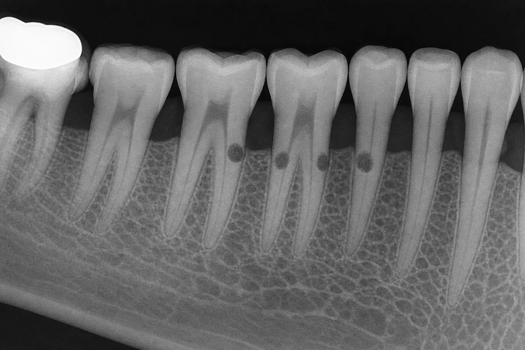
- A. Less than 1 mm
- B. Approximately 3 mmCorrect answer
- C. More than 5 mm
- D. Confined to CEJ
Answer
B. Approximately 3 mm
The radiograph shows bone levels approximately 3 mm apical to the CEJ from teeth 22 to 27.
Rationale
The periapical radiograph displays horizontal bone loss in the mandibular anterior teeth (22-27), with bone levels observed about 3 mm below the cementoenamel junction (CEJ). This is consistent with moderate chronic periodontitis. The other options are incorrect because there is clear bone loss exceeding 1 mm, not limited to the CEJ, and does not approach severe (more than 5 mm) loss.
Why each option lands where it does
- A. Underestimates the bone loss visible between teeth 22 and 27.
- B (correct). Accurately reflects the radiographic measurement of bone loss from the CEJ.
- C. Overestimates the actual bone loss; not more than 5 mm in this region.
- D. Incorrect; bone loss is not confined to the CEJ, as the crest is clearly apical.
Decision rule. Select the option that matches the radiographic measurement from the CEJ to the alveolar crest.
Takeaway. Periodontal bone loss should be measured from the CEJ, and moderate loss is typically around 3-4 mm.
Common miss. Candidates may mistake early bone loss for moderate or overlook the radiographic distance.
Patient profile 15
A 45-year-old male presents for a routine dental appointment. He reports a history of heartburn, particularly at night, and notes that his teeth have become increasingly sensitive to cold drinks over the last year. He brushes twice daily and flosses sporadically. He drinks two cups of coffee daily and frequently consumes lemon-flavored sparkling water. The patient takes omeprazole as prescribed by his physician. He does not smoke or use alcohol. There is no family history of dental problems. Examination reveals smooth, shiny enamel surfaces, especially on the palatal surfaces of the maxillary anterior teeth. Moderate plaque accumulation is noted interproximally. The patient expresses concern about his tooth sensitivity and asks for advice on how to protect his teeth. His most recent blood pressure reading is 122/78 mmHg, and he has a BMI of 26. No significant findings are observed in his extraoral examination. Radiographs demonstrate mild interproximal bone loss in the posterior maxilla and mandible.
Medical and Dental History
Medications: Omeprazole 20 mg QD (for reflux) Allergies: NKDA Social history: Non-smoker, does not drink alcohol Diet: Coffee x2/day, sparkling lemon water daily Oral hygiene: Brushes 2x/day, flosses occasionally Recent symptoms: Sensitive teeth, heartburn at night
Periodontal Chart (Teeth 3, 8, 9, 14, 19, 24, 30)
PD (mm): 3: 2/3/4 | 3/4/3 8: 2/2/2 | 2/2/3 9: 2/3/2 | 2/2/2 14: 3/4/4 | 3/4/4 19: 3/4/3 | 3/3/4 24: 2/2/2 | 2/2/3 30: 3/4/3 | 3/3/4 BOP present at sites: 3-DL, 14-MB, 19-DB, 30-ML Furcation involvement: None Mobility: All teeth normal
Clinical Findings
Enamel: Smooth, shiny surfaces on palatal aspects of 7-10, and occlusal wear on molars Plaque: Moderate interproximal accumulation Gingiva: Erythematous papillae, slight edema, no ulcerations Tooth sensitivity: Maxillary and mandibular anterior teeth
Radiographic Findings
Bitewings show mild horizontal interproximal bone loss (<2 mm from CEJ) in upper and lower posterior teeth. Anterior teeth show normal bone levels. No caries or periapical radiolucencies are visible.
Question 22
The patient requests a whitening product to address perceived yellowing of his eroded teeth. What supportive treatment service is most appropriate to provide?
- A. Recommend over-the-counter whitening strips
- B. Suggest daily hydrogen peroxide rinses
- C. Advise against whitening due to enamel lossCorrect answer
- D. Apply in-office bleaching gel
Answer
C. Advise against whitening due to enamel loss
Whitening should be discouraged in the presence of extensive enamel erosion due to risk of increased sensitivity or damage.
Rationale
Bleaching agents (both professional and OTC) can worsen sensitivity and further demineralize already eroded teeth. Daily hydrogen peroxide use can damage tissue. For patients with significant enamel loss, it's prudent to advise against whitening and instead focus on remineralization and sensitivity management.
Why each option lands where it does
- A. OTC whitening also risks exacerbating sensitivity and damage.
- B. Hydrogen peroxide rinses may harm oral tissues and are not indicated.
- C (correct). Advising against whitening protects compromised enamel and reduces sensitivity risk.
- D. In-office bleaching may worsen sensitivity and is contraindicated here.
Decision rule. Weigh cosmetic requests against clinical safety in compromised enamel.
Takeaway. Patients with erosion should avoid whitening products to prevent further damage.
Common miss. Some patients and providers underestimate the risks of bleaching in erosive cases.
Patient profile 16
A 36-year-old male presents for a dental hygiene appointment, reporting excessive gum bleeding during brushing and flossing for the past six months. He reports a history of easy bruising and prolonged bleeding after minor injuries but has not required hospitalization for his symptoms. He does not recall previous dental complications related to bleeding. His medical history lists a diagnosis of a hereditary hematologic condition managed by a hematologist. He takes prescribed medications as documented and does not smoke or use recreational drugs. The patient reports mild dental anxiety but is eager to improve his oral health, specifically requesting advice on bleeding control. On examination, heavy soft plaque deposits are visible along the gingival margins, with generalized moderate gingival inflammation. Several teeth show notable calculus accumulation. No extraoral or intraoral lesions are present. A comprehensive dental and periodontal evaluation is performed. Vital signs are within normal limits. The referring hematologist has provided guidelines for dental care in this patient.
Medication List
1. Desmopressin acetate nasal spray, 0.1 mg, as needed before procedures 2. Multivitamin with iron, daily No anticoagulants or antiplatelet agents listed.
Lab Table
Value (Patient / Reference Range) - Hemoglobin: 14.2 g/dL (13.0 – 17.5) - Platelet count: 262,000/μL (150,000 – 400,000) - Bleeding time: 11 min (3 – 8 min) - PT: 13 s (11 – 15) - aPTT: 54 s (25 – 36)
Periodontal Chart (Selected Teeth: 3, 8, 14, 19, 24, 30)
Tooth | B (mm) | MB | DB | L (mm) | ML | DL | BOP 3 | 2 | 4 | 3 | 2 | 3 | 2 | Yes 8 | 2 | 3 | 2 | 2 | 3 | 2 | Yes 14 | 3 | 4 | 3 | 3 | 4 | 3 | Yes 19 | 2 | 3 | 3 | 2 | 2 | 2 | Yes 24 | 2 | 3 | 2 | 2 | 2 | 2 | Yes 30 | 3 | 4 | 3 | 3 | 3 | 2 | Yes
Clinical Image Description
High-resolution intraoral photograph shows generalized erythematous, swollen gingiva with heavy yellow-white plaque and moderate supragingival calculus deposits on the mandibular anterior teeth.
Vital Signs
Blood Pressure: 122/76 mmHg Heart Rate: 74 bpm Respiratory Rate: 15 breaths/min Temperature: 98.4°F
Question 23
A 32-year-old patient presents with moderate gingival inflammation and no attachment loss. Which therapy is most appropriate to recommend at this stage?
- A. Daily systemic antibiotics
- B. Topical corticosteroid rinse
- C. Immediate surgical intervention
- D. Professional mechanical debridementCorrect answer
Answer
D. Professional mechanical debridement
Professional mechanical debridement is the standard of care for plaque-induced gingivitis, even in medically at-risk patients.
Rationale
Mechanical removal of plaque and calculus is the evidence-based primary therapy for gingival inflammation. Antibiotics are not indicated absent infection or advanced disease. Topical steroids are reserved for specific lesions (e.g., lichen planus), and surgery is inappropriate in this early stage and medically complex patient.
Why each option lands where it does
- A. Systemic antibiotics are not recommended for uncomplicated gingival inflammation.
- B. Topical corticosteroids are not indicated for routine plaque-induced gingivitis.
- C. Surgical intervention is not appropriate for gingivitis without periodontitis or tissue destruction.
- D (correct). Mechanical removal of plaque and calculus is the first-line treatment for gingival inflammation without attachment loss.
Decision rule. Default to mechanical therapy unless there is a clear exception.
Takeaway. Plaque removal remains first-line for gingival inflammation.
Common miss. Some may inappropriately escalate to medications or invasive care.
Patient profile 17
Mr. Brian Clark, a 58-year-old male, presents for a dental hygiene recall appointment. He reports no acute dental pain but has noticed his gums bleed during brushing and occasional tenderness in certain areas over the last several months. He reports a history of hypertension, controlled Type 2 diabetes, and hyperlipidemia. Mr. Clark states he last saw a dentist about two years ago and has recently resumed regular oral care. He quit smoking five years ago after a 20-year history (1 pack/day). He brushes once daily with a medium-bristle brush and uses mouthrinse but does not floss regularly. On examination, his blood pressure is 138/84 mmHg, and his BMI is 31. He is unemployed and reports some financial barriers to dental care. Extraoral examination is unremarkable, but intraoral inspection reveals erythmatous, swollen gingiva with areas of recession, and visible calculus. A periodontal charting and a radiograph are provided for review, along with his current medication list.
Exhibit 1: Medication List
1. Metformin 500mg BID 2. Lisinopril 20mg QD 3. Atorvastatin 20mg QD 4. Aspirin 81mg QD
Exhibit 2: Periodontal Chart (Teeth 2, 3, 14, 15, 18, 19, 30, 31)
Tooth | MB | B | DB | ML | L | DL 2 | 4 | 5 | 6 | 4 | 5 | 6 3 | 3 | 4 | 5 | 3 | 4 | 5 14 | 6 | 5 | 6 | 5 | 4 | 6 15 | 4 | 4 | 5 | 4 | 3 | 5 18 | 5 | 6 | 5 | 5 | 5 | 6 19 | 4 | 5 | 4 | 5 | 4 | 5 30 | 3 | 4 | 4 | 3 | 4 | 4 31 | 4 | 5 | 6 | 4 | 4 | 5 Furcation involvement noted on 14, 19, and 30. Bleeding on probing: 70% of sites.
Exhibit 3: Bitewing Radiograph Description
Generalized horizontal bone loss of about 30-40% on posterior teeth; vertical bone defects interproximally on teeth 14/15 and 18/19; calculus visible.
Exhibit 4: Medical History
Allergies: NKDA. Diabetes diagnosed 10 years ago, last HbA1c 7.5% (recent, per patient report). Hypertension diagnosed 5 years ago. No history of endocarditis or joint replacements. Former smoker (quit 5 years ago, 1 pack/day x 20 years).
Question 24
Based on Mr. Clark's periodontal chart and history, which factor most likely contributed to the current extent of his periodontal findings?
- A. Short-term aspirin use only
- B. Recent medium-bristle brushing
- C. Current nonsmoker status
- D. Longstanding diabetes mellitusCorrect answer
Answer
D. Longstanding diabetes mellitus
Mr. Clark’s longstanding diabetes mellitus is a major contributory factor to his periodontal findings.
Rationale
Chronic hyperglycemia impairs immune response and collagen metabolism, leading to increased susceptibility and severity of periodontal disease. Mr. Clark's HbA1c of 7.5% and 10-year diabetes duration are significant risk enhancers. While smoking is a risk, he quit five years ago, reducing its influence. Aspirin and brushing technique are minor or unrelated to the severity and distribution seen.
Why each option lands where it does
- A. Low-dose aspirin may mildly increase bleeding but does not cause periodontal destruction.
- B. Medium-bristle brushing can cause recession, but not generalized bone loss and deep pockets as seen here.
- C. His past smoking is a historical risk, but quitting five years ago has reduced its impact.
- D (correct). Diabetes, especially when not fully controlled, increases risk and severity of periodontitis by affecting tissue healing and inflammatory response.
Decision rule. Prioritize poorly controlled diabetes as a primary modifying risk factor in complex chronic periodontitis.
Takeaway. Systemic health significantly influences periodontal pathogenesis.
Common miss. Many think brushing alone is the dominant cause; in chronic cases, systemic disease is the key driver.
Patient profile 18
A 58-year-old male presents to your clinic for a routine dental hygiene appointment. He reports mild bleeding when brushing and has not seen a dentist for two years. His medical history reveals controlled hypertension and type 2 diabetes managed with medication. He is a retired construction worker who quit smoking eight years ago after a 30-pack-year history. He reports occasional dry mouth but no allergies. His last dental cleaning was about two years ago. Clinically, you note moderate plaque accumulation, gingival inflammation, and some recession. You review his current medications and collect his vital signs. A full periodontal charting and recent radiographs (taken today) are available for review.
Medication List
- Metformin 1000 mg BID - Lisinopril 20 mg QD - Hydrochlorothiazide 25 mg QD - Aspirin 81 mg QD (self-initiated) - Artificial saliva as needed
Vital Signs
BP: 138/82 mmHg Pulse: 78 bpm Resp: 15/min Temp: 98.1°F HbA1c (3 months ago): 7.1%
Periodontal Charting (Teeth #2, #3, #14, #19, #30)
Tooth D B M L MB DB 2 5 3 4 4 3 4 3 6 5 5 4 4 5 14 5 5 4 4 5 5 19 4 5 4 5 5 4 30 6 5 5 5 4 5 (Note: BOP present at 70% of sites, furcation involvement class II noted on #3 and #30) Plaque scores: 35% Gingival recession: 1-3 mm (generalized)
Bitewing Radiograph Description
The radiographs show horizontal bone loss of 20-40% in posterior regions and vertical defects at #3 and #30. Moderate calculus is visible interproximally. No carious lesions are evident.
Question 25
Examine the bitewing radiograph. Which specific interproximal area demonstrates a vertical bone defect most clearly, based on the radiographic appearance of the bone loss pattern?
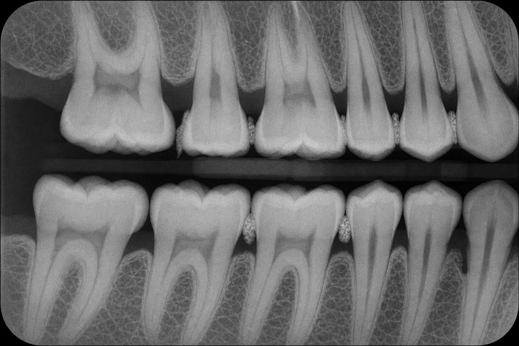
- A. Distal of #2
- B. Distal of #14
- C. Mesial of #19
- D. Mesial of #3Correct answer
Answer
D. Mesial of #3
The mesial of #3 shows a vertical bone defect, evident by the angular bone loss pattern and loss of normal crestal architecture.
Rationale
Vertical bone defects appear as angular or oblique areas of bone loss rather than the uniform reduction seen in horizontal loss. On the bitewing, the mesial of #3 displays this classic pattern, with an apex toward the root and uneven bone height between adjacent teeth. The other options show horizontal bone loss or no clear vertical component.
Why each option lands where it does
- A. Incorrect. The distal of #2 has horizontal bone loss visible, not a vertical pattern.
- B. Incorrect. The distal of #14 demonstrates horizontal, not vertical, bone loss.
- C. Incorrect. The mesial of #19 shows moderate horizontal bone loss without a vertical defect.
- D (correct). Correct. The mesial of #3 displays a vertical, angular bone defect on the radiograph.
Decision rule. Select the site that shows angular, not parallel, bone loss extending apically along the root.
Takeaway. Vertical bone defects present as angular bone loss and are best identified by their non-parallel reduction of bone height on radiographs.
Common miss. Candidates often confuse generalized horizontal loss with site-specific vertical defects, which require attention to bone contour.
Patient profile 19
A 59-year-old male presents for a dental hygiene recall appointment, reporting mild sensitivity in his lower left molars and occasional bleeding when brushing. He is a former smoker, having quit 7 years ago, and works as a truck driver. He has hypertension and Type 2 diabetes, both diagnosed over a decade ago. He reports seeing his physician regularly and checking his blood glucose daily. He brushes twice daily and uses dental floss intermittently. There are no known allergies. He expresses concern about his overall gum health but does not recall any previous periodontal treatment. He denies any recent dental trauma or facial pain. He is worried about a rough patch on his lateral tongue and requests advice about cleaning his removable partial denture. Vital signs and findings are summarized below.
Medication List
1. Metformin 1000 mg, twice daily 2. Lisinopril 20 mg, once daily 3. Atorvastatin 20 mg, once nightly 4. Over-the-counter aspirin, 81 mg, once daily
Vital Signs and Laboratory Values
Blood pressure: 132/82 mmHg Pulse: 74 bpm Respiratory rate: 14 HbA1c (last check, 4 months ago): 7.4% Fasting blood glucose (today): 134 mg/dL
Periodontal Chart (Selected Teeth: 18, 19, 20, 21, 22, 23, 24, 25, 26, 27)
All depths in mm, format: MB, B, DB / ML, L, DL 18: 5, 5, 4 / 5, 4, 4 19: 4, 5, 4 / 4, 3, 4 20: 4, 4, 4 / 3, 3, 4 21: 3, 3, 2 / 3, 2, 2 22: 3, 3, 3 / 2, 2, 3 23: 2, 2, 2 / 2, 2, 2 24: 2, 2, 2 / 2, 2, 2 25: 2, 2, 2 / 2, 2, 3 26: 3, 3, 3 / 3, 3, 3 27: 4, 4, 4 / 4, 4, 3 BOP: Sites 18MB, 19B, 19DL, 20B, 27B
Head, Neck, and Intraoral Examination
No asymmetry, swelling, or lymphadenopathy. TMJ non-tender. Palpable rough white patch, 0.7 cm, on left lateral border of tongue, non-tender, does not wipe off. Removable partial denture for teeth 29-31 present and appears clean.
Panoramic Radiograph (Described)
Generalized horizontal bone loss most prominent in posterior mandible; no periapical radiolucencies; calculus visible interproximally in mandibular molar regions; no caries evident.
Question 26
The patient mentions a friend with diabetes who skips hygiene appointments. Based on the findings, which is the most appropriate professional responsibility in this situation?
- A. Suggest patient switch physicians
- B. Educate the patient on oral-systemic linkCorrect answer
- C. Recommend only annual recall visits
- D. Dismiss the comment as nonclinical
Answer
B. Educate the patient on oral-systemic link
The best professional action is to educate the patient about the connection between diabetes and periodontal health.
Rationale
Dental hygienists have a responsibility to educate patients about health risks, especially the bidirectional relationship between diabetes and periodontal disease. This actively promotes better outcomes. Ignoring the comment, recommending changes in medical care without cause, or suggesting less frequent care are all outside best professional practice.
Why each option lands where it does
- A. Incorrect. Recommending changes in medical providers is inappropriate without evidence.
- B (correct). Correct. Patient education improves compliance and outcomes, particularly for those with chronic conditions.
- C. Incorrect. Annual visits are insufficient for high-risk patients.
- D. Incorrect. Ignoring opportunities to educate violates core responsibilities of patient advocacy and health promotion.
Decision rule. Address teachable moments with evidence-based education.
Takeaway. Professional responsibility includes patient education on systemic and oral health connections.
Common miss. Many overlook the power of brief interventions and miss opportunities for patient engagement.
Patient profile 20
A 54-year-old female presents to your dental hygiene clinic for a routine re-care appointment. She attends every six months but missed her last cleaning due to illness. She reports mild gum sensitivity during brushing and intermittent bleeding. Her medical history includes controlled hypertension and type 2 diabetes diagnosed five years ago. She occasionally experiences dry mouth. No history of tobacco use is reported. Her last dental visit was eight months ago. She brushes twice daily with a powered toothbrush and flosses most days. No known allergies. She takes her prescribed medications as directed. On clinical examination, you note generalized mild erythema, especially on the mandibular anterior gingiva. Her oral hygiene appears fair, with moderate plaque and calculus deposits. A head and neck exam is unremarkable except for mild bilateral submandibular lymph node enlargement. Periodontal charting reveals localized areas of 5 mm probing depths, with some BOP and moderate CAL. Bitewing and periapical radiographs are provided. The patient expresses concern about recurring bleeding and asks what more she can do to support her oral health.
Medical and Dental History
Age: 54 Sex: Female Medical conditions: Hypertension (controlled), Type 2 diabetes (diagnosed 5 years ago, most recent HbA1c: 7.2%) Medications: - Metformin 1000 mg BID - Lisinopril 20 mg daily Last dental visit: 8 months ago Oral hygiene: Electric toothbrush BID, flossing 5 days/week Allergies: None Tobacco use: Never
Vital Signs
Blood pressure: 128/76 mmHg Pulse: 74 bpm Respirations: 16/min Temperature: 98.3°F
Periodontal Chart (Selected)
Tooth 23: M-5mm, F-4mm, D-4mm, CAL: 4-5mm, BOP Tooth 24: M-5mm, F-4mm, D-5mm, CAL: 4-5mm, BOP Tooth 25: M-5mm, F-4mm, D-5mm, CAL: 4-5mm, BOP Generalized: 2-3mm pockets elsewhere, scattered 4mm sites, generalized slight recession Furcation: None detected
Radiographic Findings
Bitewings and periapicals show moderate horizontal bone loss in the mandibular anterior region. No caries detected. Lamina dura appears intact elsewhere.
Head and Neck Examination
TMJ: No clicks, crepitus, or limitation Facial symmetry: Normal Lymph nodes: Mild, mobile bilateral submandibular lymphadenopathy Soft tissues: No lesions or abnormalities
Question 27
No caries are evident on radiographs and no cavitated lesions are seen on oral exam. Which oral finding is most likely to be associated with her moderate dry mouth?
- A. Generalized mild mucosal erythemaCorrect answer
- B. Traumatic fibroma on tongue
- C. Leukoplakic patches on buccal mucosa
- D. Multiple aphthous ulcers
Answer
A. Generalized mild mucosal erythema
Mild mucosal erythema is often associated with dry mouth due to reduced lubrication.
Rationale
Dry mouth leads to mucosal irritation and erythema because saliva provides lubrication and antimicrobial effects. Aphthous ulcers could occur but would be found and documented if present. Leukoplakia is unrelated. Traumatic fibroma is caused by repeated trauma, not dryness.
Why each option lands where it does
- A (correct). Correct. Mild erythema aligns with the effects of reduced salivary flow.
- B. Fibromas result from chronic mechanical trauma, not xerostomia.
- C. Leukoplakia is not a sequela of dry mouth conditions.
- D. Aphthous ulcers would have been noted if present; not the most likely finding.
Decision rule. Choose the finding most commonly linked to xerostomia.
Takeaway. Mild mucosal erythema is a typical finding in mild to moderate dry mouth.
Common miss. Many overemphasize ulceration or white lesions with dry mouth.
Patient profile 21
A 54-year-old Hispanic male presents for a dental hygiene recall after 18 months without professional care. He reports occasional gum bleeding during brushing and mild tooth sensitivity to cold. His medical history reveals type 2 diabetes diagnosed 7 years ago, controlled with oral medication, and hypertension. He does not smoke or use alcohol. He reports mild dental anxiety but declines sedation. He notes dry mouth and feels tired most mornings. Recent blood work documents HbA1c at 7.9%. His last dental cleaning was before his diabetes diagnosis. He takes all medications as prescribed. The extraoral and intraoral exams are unremarkable except for dry mucosa. The following findings are available for review.
Medication List
1. Metformin 1000 mg PO BID 2. Lisinopril 20 mg PO QD 3. Hydrochlorothiazide 25 mg PO QD 4. Simvastatin 20 mg PO QHS
Vital Signs
Blood Pressure: 148/90 mmHg Heart Rate: 84 bpm Respiratory Rate: 16 BMI: 31 kg/m^2
Periodontal Chart
Probing depths (in mm): #3: 3 4 5 / 4 5 4 #8: 2 3 3 / 2 2 3 #9: 2 2 2 / 3 3 3 #14: 4 5 4 / 5 5 5 #19: 3 4 4 / 4 4 4 #24: 2 2 3 / 2 2 2 #30: 2 3 3 / 3 4 4 Bleeding on probing: Generalized, especially #14, #19 Furcation involvement: Present on #14 (Class I)
Radiographic Findings
Bitewing and periapical radiographs show moderate horizontal bone loss at maxillary and mandibular molars, widened periodontal ligament spaces on #14, and moderate interproximal calculus on posterior teeth. No periapical pathology is apparent.
Question 28
The patient rates his dental anxiety as 4/10 and is concerned about discomfort during scaling. Which evidence-based strategy is most suitable for pain management in this scenario?
- A. Topical benzocaine gelCorrect answer
- B. General anesthesia referral
- C. Behavior modification techniques
- D. Short-acting oral benzodiazepine
Answer
A. Topical benzocaine gel
Topical anesthetics minimize discomfort for mildly anxious patients.
Rationale
A moderate anxiety rating and concern about scaling pain are optimally addressed by topical anesthetic for routine procedures. Oral sedation is for more severe anxiety and requires medical clearance. General anesthesia is not warranted. Behavior modification helps but doesn't directly reduce physical pain.
Why each option lands where it does
- A (correct). Topical anesthetic is effective and safe for pain control in routine hygiene visits.
- B. General anesthesia is excessive for mild anxiety and contraindicated in most hygiene settings.
- C. Behavioral strategies reduce anxiety but not physical discomfort.
- D. Oral sedatives are not first-line for mild anxiety and carry medication interaction risks.
Decision rule. Match intervention invasiveness to anxiety severity.
Takeaway. Use topical anesthetic for mild dental anxiety and pain risk.
Common miss. There is a misconception that sedation is required for all anxious patients.
Patient profile 22
A 52-year-old male presents for a dental hygiene recall visit. He expresses concerns about recent gum bleeding when brushing and some sensitivity to cold on his lower molars. He reports brushing twice daily with a manual toothbrush but seldom flosses. He has not had a dental cleaning in over a year due to work commitments. He appears overweight and reports being recently diagnosed with type 2 diabetes. He takes prescribed medication for both diabetes and hypertension. Clinical assessment reveals heavy generalized subgingival calculus and plaque, as well as areas of gingival recession and inflammation. Radiographs are taken, and a full periodontal chart is completed. The patient also mentions stress at work and variable compliance with his medications. He is a non-smoker and denies any allergies. You review his updated medical and dental history and complete an intra- and extraoral examination. Vital signs are recorded.
Medical and Dental History
Medications: Metformin 1000 mg BID, Lisinopril 10 mg QD Medical history: Type 2 diabetes (diagnosed 1 year ago), hypertension (diagnosed 3 years ago), no allergies Last dental visit: 15 months ago Oral hygiene: Brushes twice daily; flosses 1-2x/month Social history: No tobacco or alcohol use BMI: 31 (obese)
Vital Signs
Blood pressure: 134/86 mmHg Heart rate: 76 bpm Respiratory rate: 16/min Temperature: 98.6°F
Periodontal Charting (Partial)
Teeth: 3, 14, 19, 30 Probing depths (mm): 3: MB 6, B 7, DB 6, ML 5, L 6, DL 5 14: MB 5, B 6, DB 7, ML 5, L 5, DL 6 19: MB 4, B 5, DB 5, ML 3, L 3, DL 4 30: MB 4, B 5, DB 5, ML 4, L 4, DL 3 Furcation involvement: 3 (Grade II), 14 (Grade II) Clinical attachment loss: 3-5 mm on involved teeth Bleeding on probing: Present at all sites above Gingival recession: Up to 2 mm on buccal of 3, 14
Bitewing Radiograph (Description)
Radiograph shows horizontal bone loss of 3-4 mm in molar and premolar regions bilaterally, with furcation radiolucency visible on teeth 3 and 14. Calculus deposits are radiopaque at interproximal areas.
Question 29
Refer to the bitewing radiograph. Based on the image, on which proximal surfaces is radiopaque calculus most extensively distributed among the teeth shown?
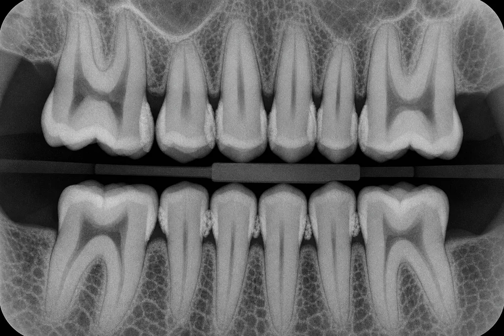
- A. Buccal of 19
- B. Mesial and distal of 3Correct answer
- C. Distal of 30
- D. Mesial of 14
Answer
B. Mesial and distal of 3
The radiograph shows pronounced radiopaque calculus deposits at both mesial and distal surfaces of tooth 3.
Rationale
Radiopaque calculus appears as discrete, irregular radiopaque areas adjacent to root surfaces on bitewing radiographs. This patient's image shows substantial calculus at both mesial and distal of tooth 3, more pronounced than at other sites. Other options either describe sites with less calculus or only a single surface. This finding is critical for risk assessment and informs instrumentation strategy.
Why each option lands where it does
- A. Buccal of 19 does not show marked radiopaque calculus in the image.
- B (correct). Keyed: Mesial and distal of 3 display dense, radiopaque calculus on the radiograph.
- C. Distal of 30 shows some calculus but much less than tooth 3.
- D. Mesial of 14 shows calculus, but not as extensively as both surfaces of 3.
Decision rule. Choose the area with the densest and most extensive calculus as seen on the radiograph.
Takeaway. Radiopaque calculus is best identified by looking for dense, irregular opacities along root surfaces—most prominent here on tooth 3.
Common miss. Candidates may focus on a single surface or misinterpret the density of calculus at other sites.
Related reading
- Periodontal assessment guide
The guide is written for the Canadian exam. Use it for the reasoning about probing and bone, and keep Universal numbers when you come back to these NBDHE items.
Frequently asked questions
Are the radiograph questions real patient films?
No. The five images are synthetic teaching exhibits made for DHPrep and labeled that way. Read the film for the finding the question asks about, then check the rationale. They are not a record of a real person.
Are the patient profiles real people?
No. The histories, charting and any teaching images are fictional material written for DHPrep. Nothing on this page is a patient record.
How much of the NBDHE is this component?
On the specifications in force, clinical services are 115 of the 200 discipline-based items. The exam also has a separate patient-case component of 150 items. The free preview is not a miniature of either form. It is 29 tagged questions, and all 29 are printed here.
Practice more with DHPrep
Create a free account to practise this component with a timer, notes, and bookmarks. The account includes the full 50-question preview. A one-time NBDHE pass opens the published US bank and the mock exams.
Drug names, prophylaxis rules and emergency steps in these items follow the references used when they were written. Check the protocol your program uses before you treat a dose or a first action as fixed.
DHPrep is an independent preparation service. These are original practice questions, not official NBDHE items, and not a prediction of a board result. DHPrep is not affiliated with, endorsed by, or sponsored by the Joint Commission on National Dental Examinations (JCNDE) or the American Dental Association (ADA).